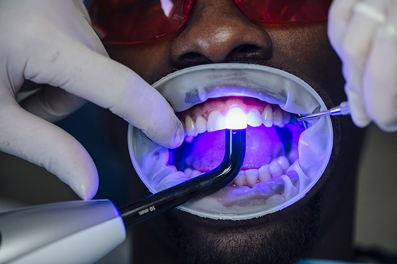
Dental imaging shows a lot to the dental providers. The x-rays let them look at the condition of the roots, teeth, jaw placement, and composition of the facial bones. The images also let them spot and treat oral problems at the initial stage of the development. In fact, x-rays can be critical in finding the problems that cannot even be noticed with an oral examination.
Treating the problems early on not just helps in saving money and a lot of pain, but also the patient’s life in some cases. With that being said, here’s outlining the top five features of dental imaging.
1. The light and dark areas of dental images
X-rays are a kind of energy that moves through or gets absorbed by the solid objects. The denser objects like the teeth and bones absorb this energy, and these show up in the x-rays as light-colored zones. On the other hand, x-rays passing through less dense objects like cheeks and gums make them come up as dark areas on the films.
2. Digital imaging for dental screening
The modern dental clinics have bid goodbye to the traditional imaging methods and embraced digital imaging. In this case, the x-rays directly reach a computer and can be seen on screen, stored, or printed. The technique is beneficial because it needs less radiation than the usual x-rays and there isn’t any wait time to develop the x-rays. The images come up on the screen within a few seconds.
The images taken of any part can be enlarged and enhanced several times of its actual size on the screen, which makes it easier for a dentist to understand exactly where and what the issue is.
3. Different types of dental imaging
There are two main types of dental imaging – intraoral (x-ray film for inside the mouth) and extraoral (x-ray film for outside the mouth). Both these types are further subdivided into several types.
For instance, intraoral x-rays are of three types, namely bitewing x-rays (upper and lower teeth in one area), periapical x-rays (the whole tooth), and occlusal x-rays (a complete tooth arch in the upper or lower jaw). Extraoral x-rays are useful in the detection of dental issues in the skull and jaw.
4. Subtraction radiography for clearer imaging
Software included in the computers can help the dental professionals run a digital comparison between current images and earlier ones through the process of subtraction radiography. Through this technique, all the things that are the same between two images get subtracted out from the image.
Thus, what is left is a clear image of the only parts that are different. It helps the dentists easily spot the smallest changes that are not noticeable to the naked eye during an oral examination.
5. The safety features of dental imaging
An extremely small amount of radiation is emitted from the x-rays. So, there is no risk of medical problems arising due to radiation. Advancements in dentistry, such as the use of x-ray machines that can limit the beam of the radiation to a smaller area, use of full-body, lead-lined aprons, and high-speed x-rays have further made the process safer for the dental professionals and the patients.
To top it off, there are federal rules and regulations that make the safety and accuracy check of x-ray machines mandatory for all the clinics.
The endnote
All the modern dental clinics are equipped with proper dental imaging tools. In fact, most clinics have moved on from the conventional tools to digital imaging techniques. As the area of digital dental imaging and reading gets more advanced, dentists will achieve more precision in their diagnosis.


